
Degenerative Disc Disease on MRI: Imaging Findings
Degenerative disc disease (DDD) is a broad term for age-related changes in the intervertebral discs, including desiccation, height loss, annular fissures, and endplate changes. While these changes are part of normal aging, symptomatic DDD can cause significant back or neck pain. MRI provides detailed evaluation of disc hydration (Pfirrmann grading), endplate changes (Modic classification), and associated findings.
Common Symptoms
- Chronic, dull axial low back or neck pain that is typically worse with prolonged sitting or standing
- Morning stiffness lasting less than 30 minutes, distinguishing it from inflammatory arthropathy
- Pain exacerbation with flexion-loading activities such as lifting and bending
- Episodic acute flares superimposed on chronic baseline pain, often triggered by minor movements
- Referred pain into the buttocks, groin, or posterior thighs without true radiculopathy
- Gradual loss of spinal height and reduced range of motion over years
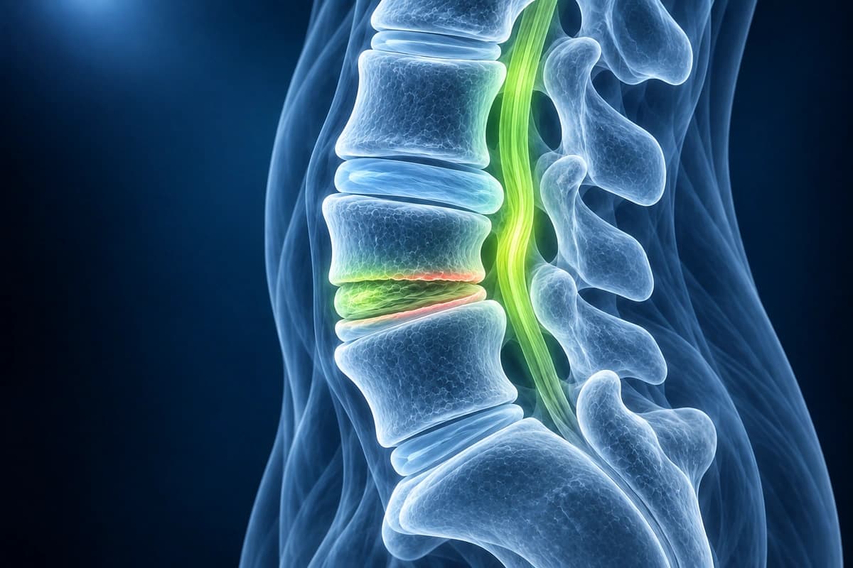
What Is Degenerative Disc Disease?
Modic Changes on a Spine MRI: Type 1, 2 and 3
Modic changes are changes in the bone marrow right next to a worn disc, where the disc meets the vertebra, and they are seen only on MRI. Type 1 looks like edema with inflammation, type 2 like fat replacing the marrow, and type 3 like hardened bone. They belong to disc degeneration, not to a separate disease.
How Modic changes appear in a report
- “Modic type 1 endplate changes at L5–S1”
- “Type 2 degenerative endplate changes at L4–L5”
- “Mixed Modic type 1/2 changes”
- “Endplate marrow edema adjacent to a degenerated disc”
Type 1, type 2 and type 3 compared
In the study that named them, type 1 was dark on T1 and bright on T2 images, and tissue samples showed fissured endplates with vascular fibrous tissue. Type 2 was bright on T1, matching fatty yellow marrow. When followed over time, type 1 turned into type 2 within 14 months to 3 years in five of six patients, while type 2 stayed stable. Type 3, dark on both, reflects sclerosis of the bone.
Do Modic changes cause back pain?
They are linked to it but do not prove it. A systematic review found Modic changes in a median of 43% of people with low back pain or sciatica and 6% of people in non-clinical groups, with an association to pain in seven of ten studies, yet some people with them have no pain. Antibiotics aimed at the finding have not held up: in a randomised trial, three months of the antibiotic amoxicillin gave no clinically important benefit over placebo for chronic back pain with Modic changes and caused more side effects.
Questions to ask about Modic changes
- Which Modic type is it, and next to which disc?
- Does that level match where my back hurts?
- Has it changed since my previous MRI?
- Which treatments do you suggest, and how strong is the evidence for them?
Annular Fissure or Annular Tear: What the Finding Means
An annular fissure is a split between the layers of fibres in the annulus, the tough outer ring of a disc. The shared spine nomenclature prefers “fissure” to “annular tear”, because the change is usually part of disc ageing rather than a sign of injury. On MRI it can show as a small bright spot called a high-intensity zone.
Ways a report can describe it
- “Posterior annular fissure at L4–L5”
- “Small posterior annular tear”
- “High-intensity zone (HIZ) in the posterior annulus”
- “Annular fissure with a small central disc protrusion”
Fissure, tear or herniation?
A fissure on its own is not a herniation. If disc material has pushed through it beyond the edge of the disc, the report names that separately as a protrusion or an extrusion, explained on the herniated disc page. Fissures are also frequent without symptoms: a review of pain-free adults found them in 19% of 20-year-olds and 29% of 80-year-olds, so the finding is weighed together with your symptoms and examination rather than treated on its own.
What to ask about an annular fissure
- Is the fissure on its own, or is there a protrusion at the same level?
- Does the report suggest a recent injury, or ageing of the disc?
- Is this level likely to be connected with my pain?
What Degenerative Disc Disease Looks Like on MRI
- Disc desiccation with loss of T2 signal (dark disc)
- Pfirrmann grading from Grade I (normal) to Grade V (collapsed disc space)
- Disc height loss at affected levels
- Annular fissures with high-intensity zone (HIZ) on T2
- Modic Type I (edema), Type II (fatty), Type III (sclerotic) endplate changes
- Osteophyte formation at vertebral body margins
Frequently Asked Questions
What does Pfirrmann grading reveal about disc degeneration?
The Pfirrmann system grades lumbar disc degeneration using routine T2-weighted MRI. Imaging changes alone do not establish the cause of pain.
What are Modic changes and why do they matter clinically?
Modic changes are MRI signal alterations in the vertebral endplates and adjacent bone marrow adjacent to degenerated discs. Type I (low T1, high T2) represents bone marrow edema and vascular granulation tissue, associated with active pain and inflammation. Type II (high T1, high T2) indicates fatty marrow conversion, generally a stable, chronic finding. Type III (low T1, low T2) represents subchondral sclerosis analogous to osteosclerosis. Conversion from type I to type II is common over time. The presence of Modic type I changes at a symptomatic level supports that level as pain-generating and guides diagnostic injections and fusion planning.
Is degenerative disc disease distinct from normal aging?
Degenerative MRI findings are common in people without back pain and become more frequent with age. Diagnosis requires clinical correlation.
Sources
- Pfirrmann CW, Metzdorf A, Zanetti M, Hodler J, Boos N. Magnetic resonance classification of lumbar intervertebral disc degeneration. Spine (Phila Pa 1976). 2001;26(17):1873-1878.
- Modic MT, Steinberg PM, Ross JS, Masaryk TJ, Carter JR. Degenerative disk disease: assessment of changes in vertebral body marrow with MR imaging. Radiology. 1988;166(1 Pt 1):193-199.
- Fardon DF, Williams AL, Dohring EJ, Murtagh FR, Gabriel Rothman SL, Sze GK. Lumbar disc nomenclature: version 2.0: recommendations of the combined task forces of the North American Spine Society, the American Society of Spine Radiology and the American Society of Neuroradiology. Spine J. 2014;14(11):2525-2545.
- Modic MT, Masaryk TJ, Ross JS, Carter JR. Imaging of degenerative disk disease. Radiology. 1988;168(1):177-186.
- Jensen TS, Karppinen J, Sorensen JS, Niinimäki J, Leboeuf-Yde C. Vertebral endplate signal changes (Modic change): a systematic literature review of prevalence and association with non-specific low back pain. Eur Spine J. 2008;17(11):1407-1422.
- Brinjikji W, Luetmer PH, Comstock B, et al. Systematic literature review of imaging features of spinal degeneration in asymptomatic populations. AJNR Am J Neuroradiol. 2015;36(4):811-816.
- Battié MC, Videman T, Parent E. Lumbar disc degeneration: epidemiology and genetic influences. Spine (Phila Pa 1976). 2004;29(23):2679-2690.
- Bråten LCH, Rolfsen MP, Espeland A, et al. Efficacy of antibiotic treatment in patients with chronic low back pain and Modic changes (the AIM study): double blind, randomised, placebo controlled, multicentre trial. BMJ. 2019;367:l5654.
Related Articles
Disc bulge vs protrusion vs extrusion vs sequestration, facet syndrome, vertebral fractures and stenosis — anatomy, MRI findings, and treatment options.
Pfirrmann grading of lumbar disc degeneration on MRI — Grade I (healthy) through V (severe collapse), what each grade means for symptoms and treatment.
Learn when 3T spine MRI can help for subtle nerve, cord, inflammatory, or post-surgical questions, and when routine 1.5T spine MRI is usually enough.
Understand your imaging before your next appointment
MRI, X-ray or CT from a CD, folder or ZIP. A PDF report or photos of the films work too.
Free to open and preview, no account needed. An optional AI report from $10 explains your images in plain language, with questions for your doctor.
Your raw files stay on this device. Only rendered images and minimal context are sent when you request an AI report.
Medical Disclaimer: This page is for informational and educational purposes only. It does not constitute medical advice, diagnosis, or treatment. AI-generated analysis may contain errors. Always consult a qualified healthcare professional for medical decisions. Full Disclaimer