
Pfirrmann Disc Grading Explained: Grade I to V on MRI
Pfirrmann grading of lumbar disc degeneration on MRI — Grade I (healthy) through V (severe collapse), what each grade means for symptoms and treatment.
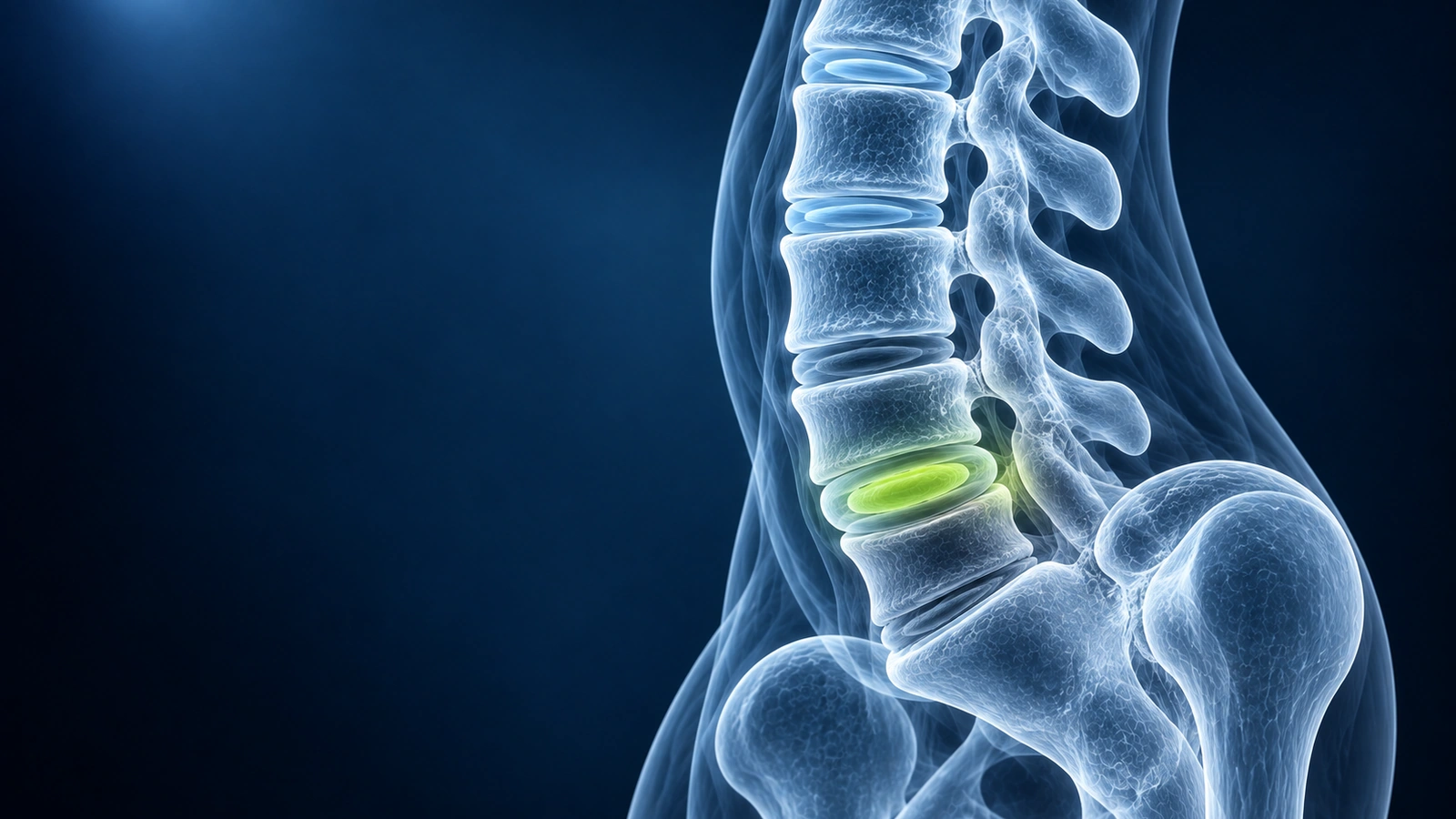
If your lumbar MRI report contains phrases like "Pfirrmann grade IV disc degeneration at L4–L5" it is natural to feel alarmed. The Pfirrmann scale is the standard radiological tool for grading how much a spinal disc has degenerated on T2-weighted sagittal MRI images, and it ranges from Grade I (a healthy, well-hydrated disc) to Grade V (a completely collapsed, desiccated disc). Understanding what each grade actually means — and crucially what it does not mean — can save you a great deal of unnecessary anxiety.
The most important fact to understand upfront: a high Pfirrmann grade does not necessarily mean you will have pain. Large population studies consistently show that 30 to 60 percent of adults with no back symptoms whatsoever have Pfirrmann grade III or IV discs on MRI. Disc degeneration is a normal part of aging — it is as universal as grey hair — and the relationship between imaging findings and pain is much weaker than most patients expect. What your spine looks like on MRI and how your spine feels are two different things.
Grade I: Healthy Disc
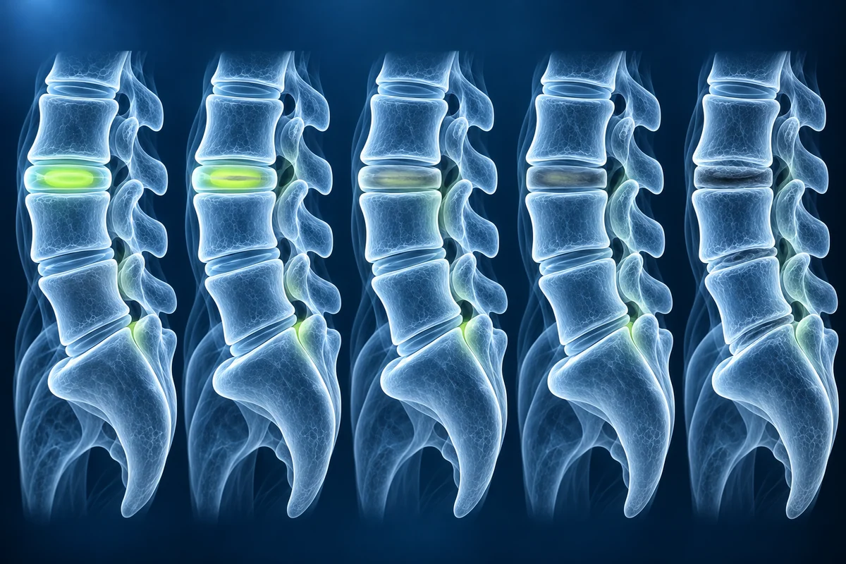
A Grade I disc is considered normal and healthy. On T2-weighted sagittal MRI the nucleus pulposus — the gel-like inner core of the disc — appears bright white, a sign that it is well-hydrated and contains a high water content. The boundary between the nucleus and the surrounding annulus fibrosus (the tough outer ring) is clearly defined and sharp. The disc height is normal for the spinal level. Grade I discs are typical in children, teenagers, and young adults, and represent the baseline against which all other grades are measured.
Grade II: Mild Degeneration
A Grade II disc still appears predominantly bright on T2 imaging, but the signal is no longer perfectly homogeneous. You may see subtle horizontal grey bands within the nucleus — these represent early fibrous tissue formation as the disc begins to lose water content. The distinction between the nucleus and annulus is still visible, and disc height remains normal. Grade II changes are extremely common in adults from their mid-twenties onwards and are generally considered within the range of normal age-related changes. The vast majority of people with Grade II discs have no symptoms related to those discs.
Grade III: Moderate Degeneration
At Grade III the nucleus has lost a significant proportion of its water content and appears as an intermediate grey signal rather than bright white on T2 imaging. The sharp boundary between the nucleus and annulus becomes blurred or indistinct, making it harder for the radiologist to identify where one ends and the other begins. Disc height is normal to slightly decreased. Grade III is the grade where patients most commonly begin to experience intermittent low back pain or stiffness — but it is equally common to find Grade III discs in completely asymptomatic people. Clinical correlation with your symptoms, physical examination, and history is essential before drawing any conclusions.
Grade IV: Severe Degeneration
Grade IV is the grade that most frequently causes patient concern, partly because it sounds severe and partly because radiologists describe it in stark terms. On T2 imaging the disc signal is dark grey to nearly black, indicating marked desiccation. There is no longer any discernible distinction between the nucleus and annulus — the disc has lost its two-compartment structure and appears as a single dark mass. Disc height is mildly to moderately reduced compared to adjacent levels. Despite the dramatic appearance, a Grade IV disc does not by itself diagnose anything, predict surgery, or mean permanent pain. Many patients with Grade IV discs at one or more levels live active, pain-free lives.
Grade IV discs do carry a higher risk of developing mechanical low back pain than Grade I or II discs, and they are more likely to be associated with disc herniations, end-plate changes, and facet arthropathy at the same level. However, the question your spine specialist will ask is not "what is the Pfirrmann grade?" but rather "does this imaging finding explain this patient's specific symptoms?" For a broader overview of disc conditions, see our page on herniated disc.
Grade V: Collapsed Disc
Grade V represents end-stage disc degeneration. The T2 signal is completely black — the disc has lost virtually all its water content. The disc space is severely or completely collapsed, and the vertebral bodies above and below may be nearly in contact. In some cases the disc space is obliterated entirely. Grade V discs are most commonly found in older adults or at levels subjected to decades of abnormal mechanical loading. At this stage the disc is essentially inert — it has little capacity to herniate because there is no fluid nuclear material remaining, and the pain generators at Grade V levels tend to be the facet joints, end-plates, and surrounding soft tissues rather than the disc itself.
Why Grade Does Not Predict Pain
This is perhaps the most clinically important concept in spine imaging, and it is supported by decades of research. A landmark 2015 study published in the American Journal of Neuroradiology found that 37 percent of asymptomatic 20-year-olds had disc degeneration on MRI, rising to 96 percent by age 80. Pfirrmann grade III or IV changes were found in 30 to 60 percent of pain-free adults across multiple large population studies. The disc is not the only pain generator in the spine — facet joints, muscles, ligaments, end-plates, and the sacroiliac joint can all produce identical symptoms. Furthermore, psychological factors, sleep quality, physical fitness, and occupational demands are often stronger predictors of disability from low back pain than imaging grade.
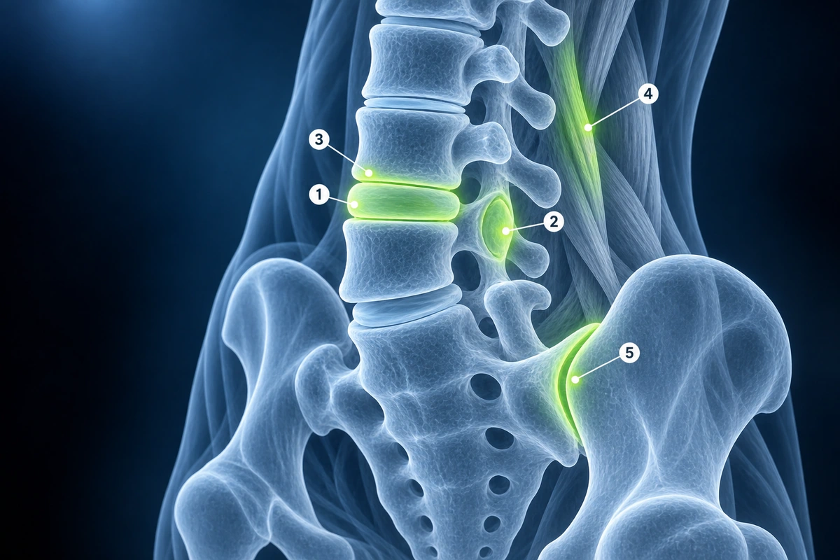
This is why spine specialists rely on clinical correlation — matching what the image shows to the patient's history, examination findings, and symptom distribution — rather than treating an MRI report in isolation. A Grade IV disc at L5–S1 in a 45-year-old marathon runner with no radicular symptoms and a normal neurological examination is very different clinically from the same imaging finding in a patient with severe S1 radiculopathy and foot weakness.
Practical Implications for Treatment
For grades I through III with no or mild symptoms, treatment is typically conservative: exercise-based rehabilitation, core strengthening, analgesics as needed, and activity modification. Grades IV and V with mechanical low back pain respond well to physical therapy focused on stabilization, load management, and aerobic conditioning. Most people with grade IV or V degeneration manage their symptoms successfully without surgery.
Surgery is rarely indicated on the basis of Pfirrmann grade alone. Surgical consideration is driven by the presence of a structural complication of degeneration — a significant disc herniation causing nerve root compression, severe spinal stenosis causing neurogenic claudication, or spondylolisthesis causing instability — not by the grade of the disc itself. Even then, surgery is generally offered only after conservative management has been given an adequate trial (typically 6 to 12 weeks), unless neurological deficits are rapidly progressive or cauda equina syndrome is present. For more on spine injury types and their treatment implications, see our guide on spine injury types explained.
Key Takeaways
- Pfirrmann grades I–V measure disc degeneration on T2 sagittal MRI based on signal intensity, nucleus-annulus distinction, and disc height
- A higher grade does not mean more pain — 30 to 60 percent of asymptomatic adults have grade III or IV discs
- Grade I is healthy; grade II shows early inhomogeneity; grade III has intermediate signal and blurred boundaries; grades IV and V are dark with height loss
- Grade V discs are end-stage but rarely the primary pain generator — facet joints and end-plates take over as pain sources at that stage
- Surgery is driven by structural complications (herniation, stenosis, instability), not by Pfirrmann grade itself
- Clinical correlation — matching imaging to your specific symptoms and examination — is always required; treat the patient, not the MRI
Frequently Asked Questions
Does a Pfirrmann grade IV disc mean I need surgery?
Almost never based on grade alone. Surgery for degenerative disc disease is indicated when a structural complication — such as a large disc herniation with nerve compression, severe central stenosis, or spondylolisthesis — causes symptoms that have not responded to a full course of conservative treatment. A Grade IV disc that is causing axial low back pain but no neurological deficit is almost always managed with physical therapy, analgesics, and activity modification first.
Can disc degeneration reverse or improve?
True reversal of degeneration (going from a higher Pfirrmann grade back to a lower one) is not possible with current non-surgical treatments. However, studies show that some discs show modest improvement in signal on repeat MRI over years, particularly with weight reduction, cessation of smoking, and regular low-impact aerobic exercise — all of which improve disc nutrition and reduce mechanical loading. More importantly, symptoms can improve substantially even when the disc appearance on MRI does not change.
Will my discs always get worse over time?
Disc degeneration is a gradual process that tends to progress slowly with age, but the rate of progression varies enormously between individuals. Genetics play a larger role than was previously appreciated — studies of identical twins show that hereditary factors account for 50 to 70 percent of the variation in disc degeneration. Modifiable factors such as obesity, heavy manual labor, smoking, and prolonged sedentary behavior accelerate progression. Maintaining a healthy weight, exercising regularly, and avoiding smoking are the most evidence-based ways to slow the process.
Should I be worried about a grade III disc finding at age 30?
Not necessarily. Grade III changes in a 30-year-old are more common than most patients realize — studies show they are present in around 30 percent of asymptomatic adults in their thirties. The finding becomes clinically relevant only if it is associated with a structural complication (herniation, stenosis) or if it correlates anatomically with your specific symptoms. An isolated Grade III disc in a young, active person with intermittent back pain and a normal neurological examination is rarely a cause for significant concern.
How does Pfirrmann grading differ from spondylosis or Modic changes?
Pfirrmann grading assesses the disc itself — its signal intensity, internal structure, and height. Spondylosis is a broader term for degenerative changes in the entire spinal motion segment, including the disc, facet joints, and osteophyte formation at the vertebral margins. Modic changes describe signal alterations in the vertebral end-plates adjacent to a degenerated disc: type I (T2-bright, T1-dark) indicates bone marrow edema and active inflammation; type II (T2-bright, T1-bright) indicates fatty end-plate conversion; type III (T2-dark, T1-dark) indicates end-plate sclerosis. Modic type I changes correlate more strongly with active back pain than Pfirrmann grade alone.
Related Articles
Decode your spine MRI report, understand disc levels, signal changes, and common findings explained in plain language.
Disc bulge vs protrusion vs extrusion vs sequestration, facet syndrome, vertebral fractures and stenosis — anatomy, MRI findings, and treatment options.
Related Conditions
Ready to analyze your imaging? Upload your MRI or X-ray for AI-powered analysis
Upload your MRI or X-ray DICOM files for private, AI-powered analysis. 4 models analyze independently — raw scan files stay in your browser.
Start AnalysisMedical Disclaimer: This page is for informational and educational purposes only. It does not constitute medical advice, diagnosis, or treatment. AI-generated analysis may contain errors. Always consult a qualified healthcare professional for medical decisions. Full Disclaimer