
Spine Injury Types Explained: Disc, Facet, and Vertebral
Disc bulge vs protrusion vs extrusion vs sequestration, facet syndrome, vertebral fractures and stenosis — anatomy, MRI findings, and treatment options.
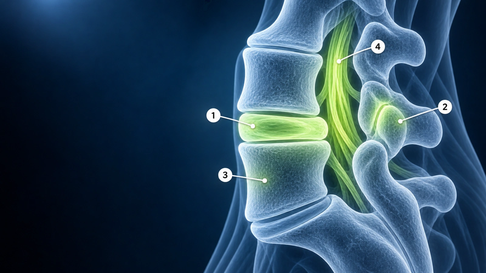
Spine injuries fall into four broad categories — disc, facet joint, vertebral, and neural canal — and the same MRI report can use terminology that means very different things clinically. Patients frequently confuse a disc bulge with a herniation, or stenosis with a herniated disc, because the terms overlap in everyday language but are distinct anatomical entities with different natural histories and treatments.
This guide uses the standardized nomenclature published by the North American Spine Society (NASS), the American Society of Neuroradiology (ASNR), and the American Society of Spine Radiology (ASSR) to clarify what each term means, how it appears on MRI, and what it generally means for treatment. For guidance on reading your own spine MRI, see how to read a spine MRI.
Disc Bulge
A disc bulge is defined as disc tissue extending beyond the vertebral endplate margins over more than 25 percent of the disc circumference (greater than 180 degrees). Broad-based extension over 25–50 percent of the circumference is called a broad-based bulge; extension over 50–100 percent is a diffuse bulge. Bulges typically represent degenerative disc disease rather than a focal mechanical event. The outer annular fibers (the disc's fibrous shell) remain intact, and the disc nucleus has not escaped its normal boundary.
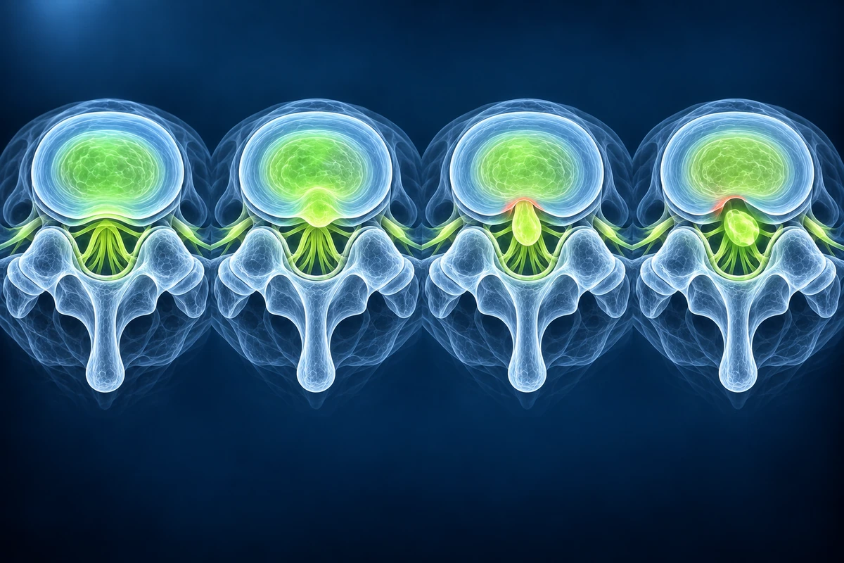
On MRI, a disc bulge appears as a smooth, symmetric or asymmetric outward convexity of the disc margin on axial images. Because a bulge involves a large arc of the disc circumference, it is less likely to compress a single nerve root than a focal protrusion or extrusion of the same height. Many disc bulges are asymptomatic incidental findings, particularly in adults over 40, where degeneration is a normal part of aging.
Disc Protrusion
A disc protrusion is a focal herniation in which the base of the displaced disc material (measured at the disc margin) is wider than the dome of the herniation in any plane. In plain terms, the disc material that has migrated out is narrower at its tip than at its origin. This distinguishes a protrusion from an extrusion (see below). Protrusions are described as focal when they involve less than 25 percent of the disc circumference, and as broad-based when they involve 25–50 percent.
On MRI, a protrusion appears as a focal convexity on axial T2-weighted images, often with T2 hypointensity (dark signal) reflecting the annular fibers still constraining the disc. Protrusions can compress the traversing or exiting nerve root and produce radicular symptoms — pain, numbness, or weakness radiating into the leg. An annular fissure (formerly called an annular tear) is a radial disruption of the annular fibers that often accompanies protrusions and appears as a focal T2 bright spot on the posterior disc margin, called a high-intensity zone (HIZ).
Disc Extrusion
A disc extrusion is a herniation in which the dome of the displaced disc material is wider than its base in at least one imaging plane — the reverse geometry of a protrusion. Imagine squeezing toothpaste through a small opening: the material that escapes is wider than the gap it came through. This occurs because the annular fibers have ruptured and the nucleus pulposus has migrated through the defect. The extruded fragment remains connected to the parent disc by a pedicle of disc material.
On MRI, an extrusion appears as a focal mass of disc signal that is clearly wider than the gap in the annulus through which it escaped. Extrusions can migrate superiorly or inferiorly along the posterior vertebral body, potentially compressing nerve roots or the thecal sac at levels adjacent to the parent disc. Extrusions are more likely to cause significant neurological symptoms than protrusions. For an overview of disc-related conditions, see our page on herniated disc.
Disc Sequestration (Free Fragment)
Sequestration is the most advanced form of disc herniation. The extruded disc fragment loses all continuity with the parent disc and becomes a free-floating fragment in the spinal canal or foramen. Because the fragment is no longer attached, it may migrate significant distances — superiorly, inferiorly, or even posteriorly — and can be found far from the disc level of origin on MRI.
On MRI, sequestered fragments often show a T2 signal different from the parent disc as they desiccate and are resorbed over time. Paradoxically, large sequestered fragments sometimes spontaneously resorb due to immune-mediated phagocytosis, which is why some patients with initially severe sciatica improve significantly over 6–12 weeks of conservative management. When neurological deficits are progressive or cauda equina syndrome is present, surgical removal is urgent.
Facet Joint Syndrome
The facet joints (also called zygapophyseal joints) are paired synovial joints at the posterior spine that guide vertebral motion and prevent excessive rotation and translation. Facet syndrome refers to pain arising from degenerative changes in these joints: loss of articular cartilage, subchondral sclerosis, osteophyte formation at the joint margins, and joint space narrowing. Facet arthropathy is extremely common with aging and is a leading source of axial low-back pain.
On MRI, facet degeneration appears as T2 hypointensity (dark signal) in the joint space reflecting cartilage loss, osteophytes as low-signal bony projections at the joint margins, and T2 hyperintensity (bright signal) in the facet joint itself when a synovial cyst or synovitis is present. Synovial cysts can extend into the lateral recess or neural foramen and compress a nerve root, mimicking disc herniation clinically. Facet-mediated pain typically worsens with extension and lateral bending, unlike discogenic pain which worsens with flexion.
Vertebral Fractures (Compression and Burst)
Vertebral fractures are classified primarily by the involvement of the posterior vertebral wall. A compression fracture involves failure of the anterior vertebral body under axial loading with resultant loss of anterior height; the posterior wall remains intact. Compression fractures are most common in osteoporotic bone and frequently occur with minimal or no trauma — a cough or rising from a chair can be sufficient. On MRI, acute compression fractures show diffuse T2 hyperintensity (bone marrow edema) and reduced T1 signal (dark) of the affected vertebra, which distinguishes acute from chronic fractures.
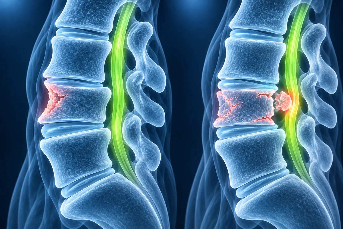
A burst fracture involves failure of both the anterior and posterior vertebral cortices, with posterior wall disruption and potential retropulsion of bone fragments into the spinal canal. Burst fractures carry a risk of spinal cord or cauda equina compression and require urgent evaluation. On MRI, posterior wall comminution and retropulsed bony fragments reducing canal diameter are the key distinguishing features from a simple compression fracture. Spinal canal compromise and neurological status guide the decision between surgical stabilization and conservative management.
Stenosis (Central, Lateral Recess, and Foraminal)
Spinal stenosis refers to narrowing of the neural spaces, and its clinical consequences depend on which space is narrowed and which neural structures are compressed. Central canal stenosis is narrowing of the central spinal canal, which contains the thecal sac, spinal cord (in the cervical and thoracic spine), or cauda equina (in the lumbar spine). Severe central stenosis causes neurogenic claudication — leg pain, heaviness, and weakness that worsen with standing and walking and improve with sitting or flexing forward.
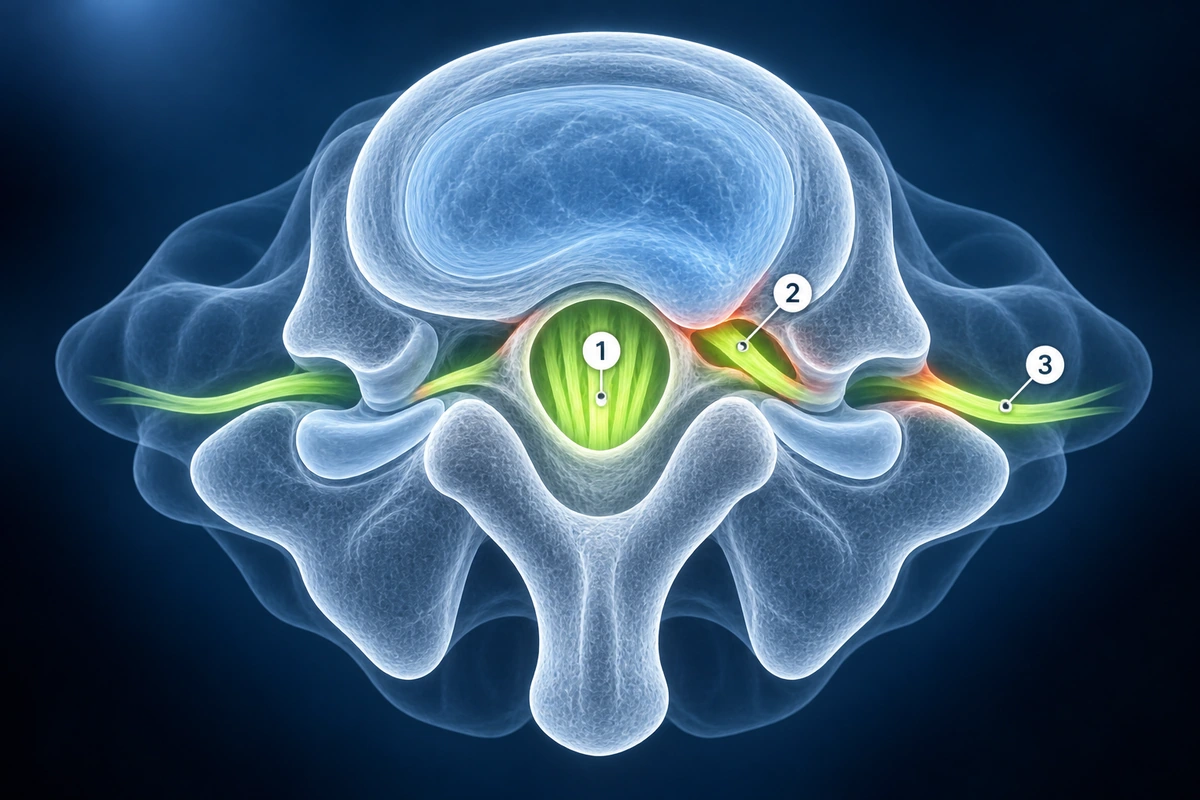
Lateral recess stenosis compresses the traversing nerve root as it travels from the thecal sac toward the foramen. In the lumbar spine, the traversing root at L4–5 is the L5 root; at L5–S1 it is the S1 root. Foraminal stenosis compresses the exiting nerve root within the neural foramen — the exiting root at L4–5 is the L4 root; at L5–S1 it is the L5 root. Distinguishing which nerve is compressed (traversing versus exiting) explains the dermatomal pattern of symptoms and guides surgical planning. Far-lateral (extraforaminal) disc herniations may compress the exiting root after it has left the foramen entirely.
Key Takeaways
- Bulge: more than 25% of the disc circumference extends beyond the endplate — broad, often degenerative, and frequently asymptomatic
- Protrusion: base wider than dome; annular fibers constrain the disc — the most common focal herniation type
- Extrusion: dome wider than base; annulus has ruptured — more likely to cause nerve compression and radicular symptoms
- Sequestration: free fragment separated from the parent disc; may spontaneously resorb but can migrate and cause cauda equina syndrome
- Facet syndrome causes extension-worsened axial pain; synovial cysts can mimic disc herniation by compressing a nerve root
- Stenosis type determines which nerve is compressed: central canal (cauda equina), lateral recess (traversing root), foramen (exiting root)
Frequently Asked Questions
What is the difference between a disc bulge and a herniation?
A disc bulge extends over more than 25 percent of the disc's circumference with the outer annular fibers intact. A herniation (protrusion or extrusion) is focal — less than 25 percent of the circumference — and involves displacement of disc material through disrupted or stretched annular fibers. Herniations are more likely to compress a specific nerve root because they concentrate disc material in a small area.
What does "broad-based" mean on a spine MRI report?
Broad-based refers to a herniation involving 25–50 percent of the disc circumference. It sits between a focal herniation (less than 25 percent) and a diffuse bulge (more than 50 percent). Broad-based herniations tend to cause more diffuse posterior pressure on the thecal sac rather than compressing a single nerve root, which is why their symptoms can be more diffuse and less radicular in character.
Do all disc findings on MRI require surgery?
No. The majority of disc findings — including bulges, protrusions, and even some extrusions — are managed conservatively with physical therapy, analgesics, and activity modification. Studies show that most radicular pain from disc herniations improves significantly within 6–12 weeks without surgery. Surgery is indicated when neurological deficits are progressive, when cauda equina syndrome is present, or when conservative treatment fails after an adequate trial.
What is an annular fissure and is it dangerous?
An annular fissure is a radial disruption of the annular fibers — the disc's fibrous outer shell. On MRI it appears as a high-intensity zone (HIZ): a focal T2-bright spot on the posterior disc margin. An annular fissure alone is not dangerous, but it represents a structural weakness that predisposes to future herniation. It can also be a source of discogenic pain if the fissure extends to the pain-sensitive outer annular fibers, even without nerve root compression.
How do stenosis symptoms differ from disc herniation symptoms?
Disc herniation typically causes acute radicular pain in one leg along a specific dermatomal distribution, worsened by flexion, coughing, or sneezing, and often improves rapidly over weeks. Stenosis (particularly central canal stenosis) causes neurogenic claudication — bilateral leg pain, heaviness, and weakness that worsen progressively with walking or standing and are relieved by sitting or leaning forward (flexing the lumbar spine opens the canal). Stenosis symptoms tend to be more chronic and gradual in onset.
Related Articles
Decode your spine MRI report, understand disc levels, signal changes, and common findings explained in plain language.
Discover the natural healing process of herniated discs, resorption rates, and when intervention is needed.
Related Conditions
Ready to analyze your imaging? Upload your MRI or X-ray for AI-powered analysis
Upload your MRI or X-ray DICOM files for private, AI-powered analysis. 4 models analyze independently — raw scan files stay in your browser.
Start AnalysisMedical Disclaimer: This page is for informational and educational purposes only. It does not constitute medical advice, diagnosis, or treatment. AI-generated analysis may contain errors. Always consult a qualified healthcare professional for medical decisions. Full Disclaimer