
Spondylolisthesis on X-ray: Imaging Findings
Spondylolisthesis is the forward displacement of one vertebral body relative to the one below. It is classified by etiology (isthmic with pars defect, degenerative, traumatic) and graded by severity using the Meyerding classification. Imaging evaluates the degree of slippage, pars integrity, disc status, and resultant neural compression.
Common Symptoms
- Low back pain that worsens with lumbar extension and activity and is relieved by flexion
- Leg pain from foraminal narrowing affecting the exiting nerve root: L4 at L4–L5 or L5 at L5–S1. Lateral recess narrowing can affect the traversing L5 or S1 root, respectively.
- Step-off deformity palpable at the spinous process level in higher-grade slips
- Hamstring tightness and spasm often causing a characteristic crouched gait in adolescent isthmic spondylolisthesis
- Neurogenic claudication in degenerative spondylolisthesis when associated spinal stenosis is present
- Bowel or bladder dysfunction as a red-flag presentation in severe high-grade slips
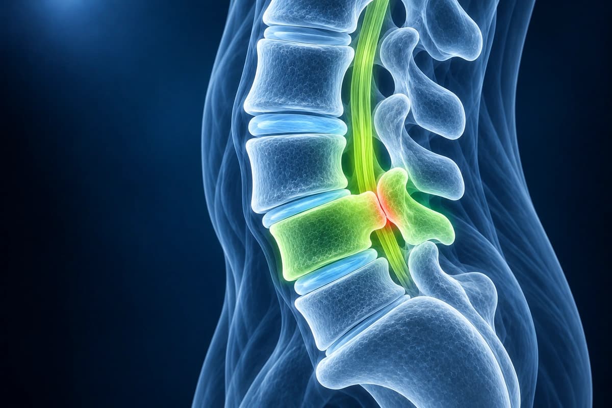
Meyerding Grades: What Grade 1, 2, 3 or 4 Spondylolisthesis Means
The Meyerding grade says how far one vertebra has slipped forward over the one below on a standing side-view X-ray. Grades divide the slip into quarters: grade 1 reaches 25%, grade 2 reaches 50%, grade 3 reaches 75% and grade 4 reaches 100%. A slip beyond 100% is grade 5, also called spondyloptosis.
How the grade is written in a report
- “Grade 1 anterolisthesis of L4 on L5”
- “Grade 1 degenerative spondylolisthesis at L4–L5 with facet arthropathy”
- “Bilateral L5 pars defects with grade 2 isthmic spondylolisthesis of L5 on S1”
- “No abnormal motion on flexion and extension views”
Low-grade and high-grade slips
Doctors group grades 1 and 2 as low grade and slips of more than 50% as high grade. High-grade slips are more often treated surgically, but the grade alone does not reliably predict symptoms or progression. Treatment also depends on the cause of the slip, spinal alignment, nerve symptoms and response to non-surgical care.
Questions about your Meyerding grade
- Which level has slipped, and is the slip degenerative or caused by a pars defect?
- Does the vertebra move further when I bend forward and back?
- Has the grade changed since my last X-ray or MRI?
- Does narrowing at this level explain my leg symptoms?
Key Imaging Findings
- Anterior translation of vertebral body relative to the one below
- Meyerding grading: Grade I (0-25%), Grade II (26-50%), Grade III (51-75%), Grade IV (76-100%)
- Pars interarticularis defect on oblique views or CT
- Associated disc degeneration at the spondylolisthesis level
- Central or foraminal stenosis from the slippage
- Reactive bone marrow changes at the pars defect
Frequently Asked Questions
What is Meyerding grading and how does it guide treatment?
The Meyerding system grades vertebral slippage on standing lateral radiographs. Management also considers alignment and dynamic stability, rather than slip grade alone.
How do MRI and CT complement each other in evaluating spondylolisthesis?
Standing lateral radiographs remain the primary tool for measuring slip percentage and assessing dynamic instability on flexion-extension views — a greater than 4 mm change in slip or greater than 10° of angular change on dynamic films confirms segmental instability. MRI best evaluates the neural consequences: foraminal stenosis compressing the exiting root, central canal narrowing, disc degeneration (Pfirrmann grading), and Modic endplate changes at the slipped level. CT with or without myelography delineates the pars interarticularis defect in isthmic spondylolisthesis (spondylolysis) and helps characterize the bony anatomy for pre-surgical planning. SPECT bone scanning is useful in adolescents to detect metabolically active stress reactions before frank pars fracture occurs.
What distinguishes isthmic from degenerative spondylolisthesis?
Isthmic spondylolisthesis results from a pars interarticularis defect (spondylolysis), typically occurring at L5–S1, and is common in adolescent athletes involved in hyperextension sports such as gymnastics and football. The slip can occur in young patients without disc degeneration. Degenerative spondylolisthesis occurs most often at L4–L5 in adults over 50, predominantly in women, and results from facet joint degeneration and disc incompetence without a pars defect — meaning the posterior elements remain intact, limiting slip to grade I–II but causing more pronounced spinal stenosis. Dysplastic spondylolisthesis is congenital and associated with sacral or L5 arch deficiency. Treatment strategy differs because degenerative cases more often require decompression for stenotic symptoms, while isthmic cases may require repair of the pars defect in younger patients.
Sources
- Wiltse LL, Newman PH, Macnab I. Classification of spondylolisis and spondylolisthesis. Clin Orthop Relat Res. 1976;(117):23-29.
- Koslosky E, Gendelberg D. Classification in brief: the Meyerding classification system of spondylolisthesis. Clin Orthop Relat Res. 2020;478(5):1125-1130.
- Daher M, Rezk A, Baroudi M, et al. Lumbar spondylolisthesis grading: current standards and important factors to consider for management. World Neurosurg. 2024;190:311-317.
- Kalichman L, Kim DH, Li L, et al. Spondylolysis and spondylolisthesis: prevalence and association with low back pain in the adult community-based population. Spine (Phila Pa 1976). 2009;34(2):199-205.
- Matz PG, Meagher RJ, Lamer T, et al. Guideline summary review: an evidence-based clinical guideline for the diagnosis and treatment of degenerative lumbar spondylolisthesis. Spine J. 2016;16(3):439-448.
- American Academy of Orthopaedic Surgeons. Spondylolysis and Spondylolisthesis. OrthoInfo.
Understand your imaging before your next appointment
MRI, X-ray or CT from a CD, folder or ZIP. A PDF report or photos of the films work too.
Free to open and preview, no account needed. An optional AI report from $10 explains your images in plain language, with questions for your doctor.
Your raw files stay on this device. Only rendered images and minimal context are sent when you request an AI report.
Medical Disclaimer: This page is for informational and educational purposes only. It does not constitute medical advice, diagnosis, or treatment. AI-generated analysis may contain errors. Always consult a qualified healthcare professional for medical decisions. Full Disclaimer