
Spinal Cord Injury on MRI & X-ray: Imaging Findings
Spinal cord injury encompasses traumatic and non-traumatic damage to the spinal cord that can result in motor, sensory, and autonomic dysfunction below the level of injury. MRI is the definitive imaging modality for evaluating cord pathology, detecting intramedullary signal abnormalities, hemorrhage, edema, and cord compression. The extent of cord signal change on MRI correlates with neurological prognosis.
Some signs described on this page need prompt medical care. If you have them, get care first. Reading about imaging can wait.
Common Symptoms
- Complete or partial loss of motor function below the neurological level of injury, ranging from mild weakness to complete paralysis
- Loss of sensation including pain, temperature, touch, and proprioception below the level of injury in a pattern depending on injury completeness and cord syndrome
- Autonomic dysfunction including neurogenic bladder (typically spastic or flaccid), bowel dysfunction, and sexual dysfunction
- Neuropathic pain described as burning, electric, or stabbing, arising from the injury level or in denervated regions
- Spasticity of limbs below the lesion level in upper motor neuron injuries due to loss of descending inhibition
- Autonomic dysreflexia in injuries above T6 — sudden severe hypertension triggered by noxious stimuli below the lesion, presenting as headache, sweating, and bradycardia
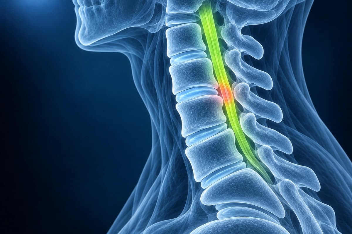
What We Look For on Imaging
- Intramedullary T2 hyperintensity indicating cord edema or myelomalacia
- T1 hypointensity and T2 signal void within the cord suggesting hemorrhage
- Cord compression from disc herniation, fracture fragment, or epidural hematoma
- Spinal canal stenosis with measurement of canal compromise percentage
- Ligamentous injury including disruption of the posterior ligamentous complex
- Cord swelling, atrophy, or syrinx formation as sequelae of injury
Frequently Asked Questions
How is spinal cord injury classified using the ASIA Impairment Scale?
The American Spinal Injury Association (ASIA) Impairment Scale (AIS) classifies injury severity based on the International Standards for Neurological Classification of SCI (ISNCSCI). AIS A (complete) indicates no motor or sensory function preserved in sacral segments S4–S5. AIS B (sensory incomplete) preserves sensation but not motor function below the neurological level including S4–S5. AIS C (motor incomplete) preserves motor function below the level with more than half of key muscles having a grade less than 3. AIS D (motor incomplete) means more than half of key muscles below the level have grade 3 or better. AIS E indicates normal motor and sensory function throughout. The neurological level of injury (NLI) is the most caudal level with intact motor and sensory function bilaterally. Sacral sparing (preserved perianal sensation or voluntary anal contraction) is the critical distinction between complete and incomplete injury.
What MRI findings are most prognostic after acute spinal cord injury?
MRI helps assess acute spinal cord injury and prognosis. Normal cord signal, single-level edema, multilevel edema, and combined hemorrhage and edema have different prognostic implications.
What are the immediate management priorities after acute spinal cord injury, and when is surgery indicated?
In the observational STASCIS study, at least two AIS grades of improvement at six months occurred in 19.8% of patients decompressed within 24 hours and 8.8% treated later.
Sources
- Fehlings MG, Vaccaro A, Wilson JR, et al. Early versus delayed decompression for traumatic cervical spinal cord injury: results of the Surgical Timing in Acute Spinal Cord Injury Study (STASCIS). PLoS One. 2012;7(2):e32037.
- Bozzo A, Marcoux J, Radhakrishna M, et al. The role of magnetic resonance imaging in the management of acute spinal cord injury. J Neurotrauma. 2011;28(8):1401-1411.
- Expert Panel on Neurological Imaging and Musculoskeletal Imaging; Beckmann NM, West OC, et al. ACR Appropriateness Criteria Suspected Spine Trauma. J Am Coll Radiol. 2019;16(5S):S264-S285.
Do not delay urgent medical care to use this tool.
After you get care, understand your imaging
MRI, X-ray or CT from a CD, folder or ZIP. A PDF report or photos of the films work too.
Free to open and preview, no account needed. An optional AI report from $10 explains your images in plain language, with questions for your doctor.
Your raw files stay on this device. Only rendered images and minimal context are sent when you request an AI report.
Medical Disclaimer: This page is for informational and educational purposes only. It does not constitute medical advice, diagnosis, or treatment. AI-generated analysis may contain errors. Always consult a qualified healthcare professional for medical decisions. Full Disclaimer