
Common Symptoms
- Saddle anesthesia — numbness or tingling around the genitals, buttocks, and inner thighs in the distribution of sacral nerve roots S3 to S5
- New-onset urinary retention — inability to initiate urination — or urinary incontinence without previous history
- Fecal incontinence or loss of voluntary bowel control
- Bilateral leg weakness or numbness, often asymmetric in early presentation
- Severe low back pain with bilateral radicular pain radiating into both legs
- Sexual dysfunction including reduced perineal sensation or genital numbness
Cauda Equina Syndrome on MRI: AI-Powered Imaging Analysis
AI-powered cauda equina syndrome imaging analysis on lumbar MRI. Saddle anesthesia, urinary retention, large central disc herniation — a serious condition best evaluated promptly by a clinician.
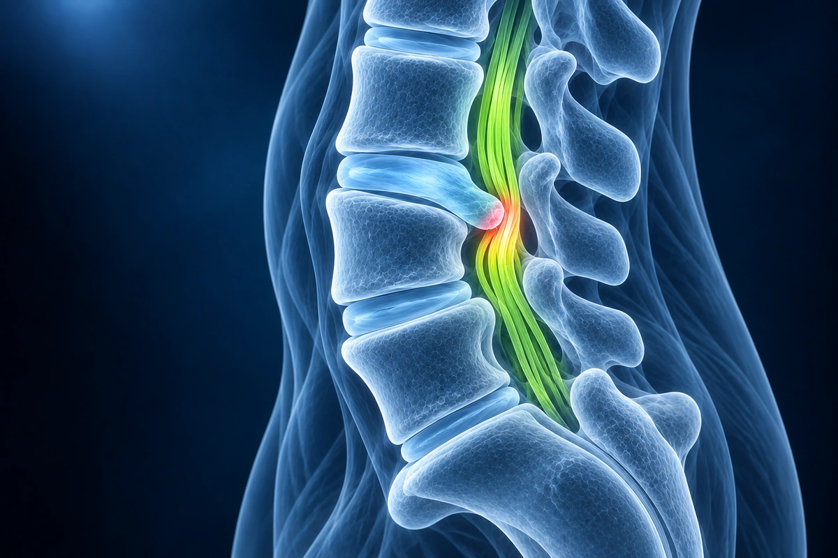
Cauda equina syndrome is a surgical emergency. If you have new-onset saddle numbness — numbness around the genitals, buttocks, or inner thighs — difficulty urinating, or fecal incontinence alongside back pain, go to the emergency department immediately. Do not wait for a routine appointment.
Surgical decompression within 48 hours of symptom onset dramatically improves neurological outcomes, including recovery of bladder and bowel control. Delays beyond 48 hours are associated with significantly higher rates of permanent sphincteric dysfunction, saddle numbness, and lower-limb weakness.
The condition results from compression of the cauda equina — the bundle of nerve roots that descend from the spinal cord below the L1 vertebra, supplying sensation and motor function to the legs, bladder, bowel, and sexual organs. The most common cause is a large central disc herniation, but cauda equina syndrome can also result from a spinal epidural abscess, epidural hematoma, spinal tumor, or severe traumatic vertebral burst fracture. MRI of the lumbar spine is the definitive imaging investigation and is performed on an emergency basis when cauda equina syndrome is suspected.
What We Look For on Imaging
- Large central or paracentral disc herniation completely or near-completely effacing the thecal sac on sagittal T2-weighted images
- Cauda equina nerve root compression or crowding on axial T2 images at the level of herniation
- Displaced or clumped nerve roots within the thecal sac below the level of compression
- Epidural mass, abscess, or hematoma with rim enhancement on post-contrast sequences when infection or vascular cause is suspected
- Post-traumatic vertebral burst fracture with posterior wall retropulsion into the spinal canal
- Severe multilevel central canal stenosis with acute decompensation, often with superimposed disc herniation or epidural lipomatosis
Frequently Asked Questions
How quickly do I need surgery if cauda equina syndrome is confirmed?
The goal is surgical decompression within 48 hours from the onset of sphincteric symptoms — urinary retention, incontinence, or fecal incontinence. Most spine surgeons aim for emergency decompression within 24 hours when possible. Patients who undergo surgery within 48 hours have significantly better rates of bladder and bowel recovery than those who are operated on later. If MRI confirms cauda equina compression, the surgical team will not wait for a routine operating slot — this is treated as an emergency alongside trauma and vascular emergencies.
Can cauda equina syndrome be diagnosed without an MRI?
Clinical examination can raise strong suspicion — saddle anesthesia, absent anal tone, urinary retention, and bilateral leg weakness in a patient with severe back pain is a classic presentation. However, MRI is required to confirm the diagnosis, identify the exact level and cause of compression, rule out alternative diagnoses such as epidural abscess, and guide surgical planning. CT myelography is used when MRI is contraindicated (for example in patients with certain pacemakers). Plain X-rays are not sufficient to diagnose cauda equina syndrome.
Will I recover fully after surgery for cauda equina syndrome?
Recovery depends primarily on how severe the compression was, how long symptoms were present before decompression, and the patient's baseline neurological status at the time of surgery. Leg pain and weakness generally have the best recovery — most patients see significant improvement in radicular symptoms within weeks to months of surgery. Bladder function recovery is less predictable: patients with urinary retention who undergo surgery within 48 hours recover continence in the majority of cases, though recovery can take months and may be incomplete. Bowel and sexual function recovery tends to lag behind motor and sensory recovery. Saddle numbness can persist for months even after successful decompression. Incomplete cauda equina syndrome — where some function is preserved at presentation — has a better prognosis than complete syndrome where all sacral functions are lost.
Upload your imaging to check for Cauda Equina
Upload your MRI or X-ray DICOM files for private, AI-powered analysis. 4 models analyze independently — raw scan files stay in your browser.
Upload & AnalyzeMedical Disclaimer: This page is for informational and educational purposes only. It does not constitute medical advice, diagnosis, or treatment. AI-generated analysis may contain errors. Always consult a qualified healthcare professional for medical decisions. Full Disclaimer