
Compression Fracture on X-ray: Imaging Findings
Vertebral compression fractures (VCFs) are the most common osteoporotic fractures, affecting approximately 25% of postmenopausal women. MRI is critical for determining fracture acuity (acute vs. chronic), differentiating benign osteoporotic fractures from pathological fractures due to malignancy, and identifying complications such as retropulsion and cord compression.
Common Symptoms
- Sudden onset of severe, localized axial back pain typically following minimal trauma in osteoporotic patients
- Point tenderness directly over the fractured vertebral level on midline percussion
- Pain worsening with upright loading and weight-bearing, improving with supine rest
- Height loss and progressive kyphotic deformity (dowager's hump) with multiple chronic fractures
- Radicular pain or neurological deficit when retropulsed bone fragments compromise the spinal canal or neural foramen
- Referred abdominal or chest pain in thoracic fractures mimicking visceral pathology
Key Imaging Findings
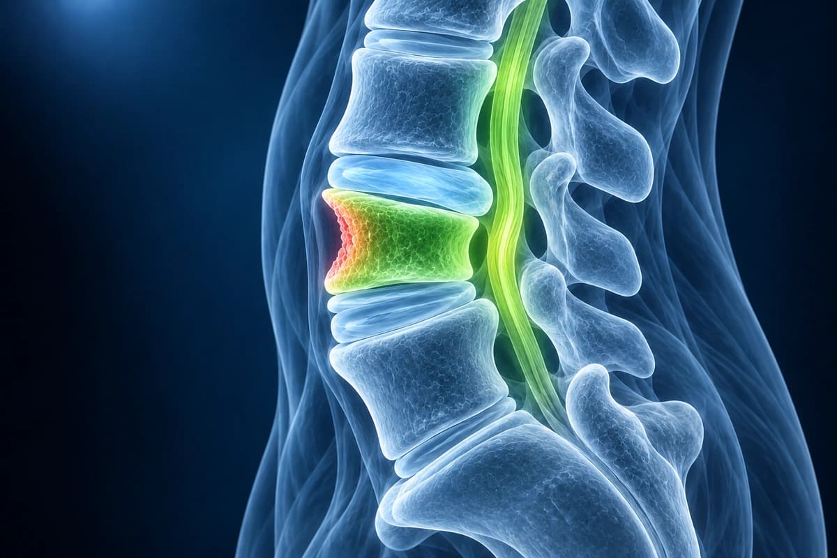
- Vertebral body height loss with wedge, biconcave, or crush morphology
- Bone marrow edema on MRI (STIR/T2 fat-sat) indicating acute fracture
- Absence of marrow edema suggesting chronic/healed fracture
- Retropulsion of fracture fragment into the spinal canal
- Convex posterior vertebral body contour suspicious for malignancy
- Enhancement pattern helping differentiate benign from pathological fracture
Frequently Asked Questions
How does MRI distinguish acute from chronic compression fractures?
Acute and subacute compression fractures show marrow edema on MRI — low signal on T1 and high signal on STIR (short tau inversion recovery) sequences — reflecting the bone marrow edema phase that persists for weeks to months. Chronic fractures have normalized marrow signal isointense to adjacent vertebrae on both sequences. The distinction is clinically critical because acute fractures may respond to percutaneous vertebroplasty or kyphoplasty within 6–8 weeks of onset, while chronic fractures do not benefit. Gadolinium enhancement patterns also differ: benign fractures show diffuse or bandlike enhancement, whereas pathological fractures from metastatic disease or myeloma show heterogeneous nodular enhancement and often convex posterior cortex bulging.
What classification systems are used for thoracolumbar fractures?
Treatment decisions consider fracture morphology, posterior ligament integrity, and neurological status. A two-column rule alone does not determine whether surgery is needed.
When should compression fractures prompt investigation for underlying pathology?
Fragility fractures occurring with minimal or no trauma in patients over 50 should trigger dual-energy X-ray absorptiometry (DXA) to assess bone mineral density and guide osteoporosis treatment. Red flags suggesting pathological fracture from malignancy include fracture above T4, multiple non-contiguous fractures, abnormal soft-tissue mass, convex posterior cortex, pedicle destruction visible on CT, age under 55 without osteoporosis, or a history of primary malignancy. MRI with gadolinium or CT-guided biopsy is required when malignancy is suspected. Myeloma should be excluded with serum and urine protein electrophoresis, immunofixation, and free light chain assay in any patient over 50 with vertebral collapse and an elevated erythrocyte sedimentation rate.
Sources
- Alexandru D, So W. Evaluation and management of vertebral compression fractures. Perm J. 2012;16(4):46-51.
- Genant HK, Wu CY, van Kuijk C, Nevitt MC. Vertebral fracture assessment using a semiquantitative technique. J Bone Miner Res. 1993;8(9):1137-1148.
- Denis F. The three column spine and its significance in the classification of acute thoracolumbar spinal injuries. Spine (Phila Pa 1976). 1983;8(8):817-831.
- Vaccaro AR, Lehman RA Jr, Hurlbert RJ, et al. A new classification of thoracolumbar injuries: the importance of injury morphology, the integrity of the posterior ligamentous complex, and neurologic status. Spine (Phila Pa 1976). 2005;30(20):2325-2333.
- Jung HS, Jee WH, McCauley TR, et al. Discrimination of metastatic from acute osteoporotic compression spinal fractures with MR imaging. Radiographics. 2003;23(1):179-187.
Related Articles
Compare CT and MRI scans, their strengths for different conditions, radiation exposure, cost, and when each is the better choice.
Decode your spine MRI report, understand disc levels, signal changes, and common findings explained in plain language.
Understand your imaging before your next appointment
MRI, X-ray or CT from a CD, folder or ZIP. A PDF report or photos of the films work too.
Free to open and preview, no account needed. An optional AI report from $10 explains your images in plain language, with questions for your doctor.
Your raw files stay on this device. Only rendered images and minimal context are sent when you request an AI report.
Medical Disclaimer: This page is for informational and educational purposes only. It does not constitute medical advice, diagnosis, or treatment. AI-generated analysis may contain errors. Always consult a qualified healthcare professional for medical decisions. Full Disclaimer