
Ankylosing Spondylitis MRI Signs: Sacroiliitis, Romanus Lesions, and Bamboo Spine
Ankylosing spondylitis is a chronic inflammatory arthritis primarily affecting the axial skeleton, with sacroiliitis as its hallmark feature. The disease typically begins in young adults and is strongly associated with HLA-B27. Progressive inflammation leads to syndesmophyte formation, ligamentous ossification, and eventual spinal fusion (bamboo spine). MRI is the most sensitive modality for detecting early sacroiliitis before radiographic changes appear. AI can help explain report terms about inflammation and structural changes, but a clinician must confirm the diagnosis and monitor the condition.
Common Symptoms
- Insidious onset of chronic inflammatory low back pain before age 40, lasting more than 3 months, improving with exercise and not relieved by rest
- Morning stiffness lasting more than 60 minutes that improves with movement and heat
- Alternating buttock pain due to sacroiliitis, characteristically switching sides
- Reduced chest expansion (less than 2.5 cm at the 4th intercostal level) and restricted spinal mobility measured by the Schober test
- Enthesitis at the Achilles tendon insertions, plantar fascia, and pelvic brim causing heel pain and rib pain
- Extra-articular manifestations including anterior uveitis (eye redness and photophobia), psoriasis, and inflammatory bowel disease
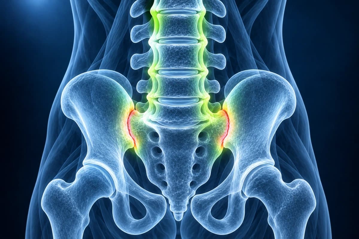
What Is Ankylosing Spondylitis?
MRI is strongest for active inflammatory lesions such as sacroiliac bone marrow edema and vertebral corner inflammation, while CT and X-ray better show chronic structural change once syndesmophytes or ankylosis are present. If symptoms look more mechanical than inflammatory, compare with herniated disc and facet joint syndrome.
Red flags are different in an ankylosed spine: new pain after even minor trauma can represent an unstable fracture, and neurological symptoms need urgent medical review. Use the free spine MRI viewer to inspect sequences locally, then share concerning studies with a radiologist or rheumatology team for diagnostic correlation and treatment decisions.
Can MRI Show Ankylosing Spondylitis Before an X-ray Does?
Often, yes. MRI can show active inflammation in the sacroiliac joints, seen as bone marrow edema under the joint surface, before X-rays show any damage. That is why the ASAS criteria accept active sacroiliitis on MRI for classifying axial spondyloarthritis, the wider group that includes ankylosing spondylitis. A rheumatologist still has to judge whether the inflammation fits the disease.
Wording on a sacroiliac joint MRI report
- “Subchondral bone marrow edema on the iliac side of the left sacroiliac joint”
- “Findings consistent with active sacroiliitis”
- “Erosions and periarticular fat metaplasia”
- “No active inflammatory change of the sacroiliac joints”
The ASAS group still treats clear bone marrow edema in the bone next to the joint as the defining sign of active sacroiliitis. Erosions or fat metaplasia support the reading but are not required. X-rays remain part of the older modified New York criteria, which define ankylosing spondylitis by definite sacroiliitis on X-ray together with clinical features such as inflammatory back pain or reduced spinal movement.
Ankylosing Spondylitis on MRI and X-ray: Key Signs
- Sacroiliac joint bone marrow edema on STIR sequences indicating active inflammation
- Erosions and sclerosis along the sacroiliac joint surfaces
- Syndesmophytes bridging adjacent vertebral bodies along the anterior longitudinal ligament
- Corner inflammatory lesions (Romanus lesions) at the vertebral body margins
- Fat metaplasia (fatty Romanus lesions) indicating prior inflammation
- Progressive ankylosis with sacroiliac joint fusion and spinal bamboo spine appearance
- CT-visible fracture lines or posterior element extension after trauma in an ankylosed spine
Frequently Asked Questions
What MRI findings diagnose early axial spondyloarthritis before radiographic changes appear?
The Assessment of SpondyloArthritis International Society (ASAS) definition of active sacroiliitis on MRI requires bone marrow edema (osteitis) on STIR or T1 fat-saturated post-gadolinium sequences in a typical sacroiliac joint distribution — specifically in the subchondral bone of the iliac or sacral side of the joint. A single clearly positive quadrant on two consecutive slices, or two positive quadrants on a single slice, meets the definition. MRI can detect sacroiliitis years before structural damage becomes visible on plain radiographs, enabling early diagnosis in the non-radiographic axial spondyloarthritis (nr-axSpA) phase and initiation of biologic therapy. Structural lesions (fat metaplasia, erosions, sclerosis, ankylosis) represent chronic damage and are better seen on CT.
How does ankylosing spondylitis affect the spine radiographically and what is the bamboo spine?
Spinal involvement in AS progresses from inflammatory erosion of vertebral corner Romanus lesions (shiny corner sign on MRI STIR or T1 gadolinium) to fatty change at the vertebral corners (fatty Romanus lesions), then to syndesmophyte formation (vertical bony bridging of the annulus fibrosus) and ultimately complete intervertebral fusion. The bamboo spine describes complete fusion of the vertebral column with continuous vertically oriented syndesmophytes on AP radiograph, accompanied by fusion of the apophyseal joints (trolley-track sign) and posterior spinal ligament ossification. This rigid column is highly susceptible to fracture — even low-energy trauma can cause unstable three-column fractures, most commonly at C5–C7, which carry high risk of neurological deficit and must be evaluated with full-spine CT and MRI regardless of symptom severity.
What is the role of HLA-B27 and biologics in management?
ASAS axial spondyloarthritis classification requires sacroiliitis on imaging plus at least one spondyloarthritis feature, or HLA-B27 positivity plus at least two features. Clinical interpretation remains essential.
Sources
- van der Linden S, Valkenburg HA, Cats A. Evaluation of diagnostic criteria for ankylosing spondylitis. A proposal for modification of the New York criteria. Arthritis Rheum. 1984;27(4):361-368.
- Rudwaleit M, van der Heijde D, Landewé R, et al. The development of Assessment of SpondyloArthritis international Society classification criteria for axial spondyloarthritis (part II): validation and final selection. Ann Rheum Dis. 2009;68(6):777-783.
- Lambert RG, Bakker PA, van der Heijde D, et al. Defining active sacroiliitis on MRI for classification of axial spondyloarthritis: update by the ASAS MRI working group. Ann Rheum Dis. 2016;75(11):1958-1963.
- Rudwaleit M, Jurik AG, Hermann KG, et al. Defining active sacroiliitis on magnetic resonance imaging (MRI) for classification of axial spondyloarthritis: a consensual approach by the ASAS/OMERACT MRI group. Ann Rheum Dis. 2009;68(10):1520-1527.
- Sieper J, Poddubnyy D. Axial spondyloarthritis. Lancet. 2017;390(10089):73-84.
- Ramiro S, Nikiphorou E, Sepriano A, et al. ASAS-EULAR recommendations for the management of axial spondyloarthritis: 2022 update. Ann Rheum Dis. 2023;82(1):19-34.
Understand your imaging before your next appointment
MRI, X-ray or CT from a CD, folder or ZIP. A PDF report or photos of the films work too.
Free to open and preview, no account needed. An optional AI report from $10 explains your images in plain language, with questions for your doctor.
Your raw files stay on this device. Only rendered images and minimal context are sent when you request an AI report.
Medical Disclaimer: This page is for informational and educational purposes only. It does not constitute medical advice, diagnosis, or treatment. AI-generated analysis may contain errors. Always consult a qualified healthcare professional for medical decisions. Full Disclaimer