
Scoliosis on MRI & X-ray: Imaging Findings
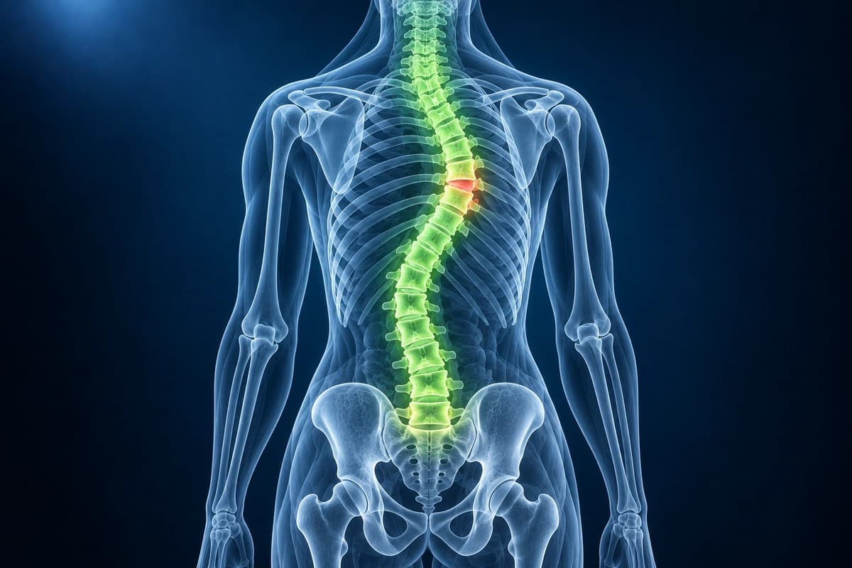
Scoliosis is an abnormal lateral curvature of the spine measuring 10 degrees or more by Cobb angle. It is classified as structural (fixed vertebral rotation) or functional (correctable postural curvature). Adolescent idiopathic scoliosis is the most common form, while degenerative scoliosis develops in adults from asymmetric disc and facet degeneration. Imaging is essential for measuring curve magnitude, monitoring progression, and identifying underlying causes such as congenital vertebral anomalies or intraspinal pathology.
Common Symptoms
- Visible lateral curvature of the spine with shoulder asymmetry, uneven waistline, or one hip appearing higher than the other
- Cosmetic trunk asymmetry including rib hump (thoracic rotation) visible on Adams forward bend test
- Back pain in adult scoliosis, often mechanical in nature, worsened by prolonged standing and relieved by rest
- Radicular leg pain in degenerative (de novo) adult scoliosis due to asymmetric foraminal stenosis on the concave side
- Reduced pulmonary function and exertional dyspnea when thoracic Cobb angle exceeds 70–80° due to restrictive chest wall deformity
- Progressive neurological symptoms including weakness or bowel and bladder dysfunction in severe adult scoliosis with associated stenosis
What We Look For on Imaging
- Cobb angle measurement to quantify the magnitude of the primary and compensatory curves
- Vertebral rotation and pedicle asymmetry indicating structural scoliosis
- Apical vertebra identification and curve classification (King, Lenke systems)
- Skeletal maturity assessment using Risser sign for adolescent growth prediction
- Intraspinal anomalies such as syringomyelia, Chiari malformation, or tethered cord
- Degenerative changes including disc space narrowing and lateral listhesis in adult curves
Frequently Asked Questions
What is the Cobb angle and how does it determine treatment?
The Cobb angle is measured on a full-length standing posteroanterior (PA) radiograph by drawing lines along the endplates of the most tilted vertebrae at the upper and lower ends of the curve — the angle between these lines (or their perpendiculars) is the Cobb angle. Curves less than 10° are considered normal spinal variation; 10–24° are monitored with serial radiographs every 6–12 months in skeletally immature patients (Risser 0–2). Curves of 25–40° with documented progression of greater than 5° are managed with thoracolumbosacral orthosis (TLSO) bracing to prevent further progression until skeletal maturity. Curves exceeding 45–50° in adolescents, or greater than 50° in adults with pain or neurological compromise, are considered for surgical correction with instrumented spinal fusion. EOS low-dose biplanar imaging minimizes cumulative radiation in young patients requiring serial monitoring.
How does MRI contribute to scoliosis evaluation beyond the Cobb angle?
Standing radiographs define curve magnitude and skeletal maturity (Risser sign of iliac apophysis ossification), but MRI is indicated when curve patterns are atypical or neurological symptoms are present. Left thoracic curves in adolescents and rapidly progressive curves are associated with underlying spinal cord abnormalities — Chiari malformation type I, syringomyelia, tethered cord, or spinal cord tumor — that can drive curve progression and must be excluded before bracing or surgery. In adult degenerative scoliosis, MRI assesses the degree of disc degeneration (Pfirrmann grading), facet arthropathy, Modic endplate changes, foraminal and central canal stenosis, and coronal imbalance — all critical for surgical planning including the need for interbody reconstruction and extent of instrumentation.
What distinguishes idiopathic adolescent scoliosis from adult de novo scoliosis?
Adolescent idiopathic scoliosis (AIS) develops during the growth spurt (ages 10–16), has no identifiable cause, and is most common in girls. It is typically asymptomatic initially, detected on school screening or by parental observation. The natural history depends on curve magnitude at skeletal maturity — curves below 30° at maturity rarely progress, while curves above 50° tend to progress at approximately 1° per year throughout life. Adult de novo scoliosis develops after skeletal maturity due to asymmetric degenerative disc disease and facet arthropathy, most often in women after age 50 and frequently presenting with back pain, lateral listhesis, and neurogenic claudication. Distinguishing the two is important because treatment goals differ: AIS aims to prevent progression and cosmetic deformity, whereas adult scoliosis management primarily targets pain and neurological function.
Sources
- Weinstein SL, Dolan LA, Cheng JC, et al. Adolescent idiopathic scoliosis. Lancet. 2008;371(9623):1527-1537.
- Lenke LG, Betz RR, Harms J, et al. Adolescent idiopathic scoliosis: a new classification to determine extent of spinal arthrodesis. J Bone Joint Surg Am. 2001;83(8):1169-1181.
- Weinstein SL, Dolan LA, Wright JG, Dobbs MB. Effects of bracing in adolescents with idiopathic scoliosis. N Engl J Med. 2013;369(16):1512-1521.
- Aebi M. The adult scoliosis. Eur Spine J. 2005;14(10):925-948.
Understand your imaging before your next appointment
MRI, X-ray or CT from a CD, folder or ZIP. A PDF report or photos of the films work too.
Free to open and preview, no account needed. An optional AI report from $10 explains your images in plain language, with questions for your doctor.
Your raw files stay on this device. Only rendered images and minimal context are sent when you request an AI report.
Medical Disclaimer: This page is for informational and educational purposes only. It does not constitute medical advice, diagnosis, or treatment. AI-generated analysis may contain errors. Always consult a qualified healthcare professional for medical decisions. Full Disclaimer