Spinal Stenosis on MRI: Imaging Findings
Spinal stenosis is the narrowing of the spinal canal, lateral recesses, or neural foramina, resulting in compression of the spinal cord or nerve roots. It most commonly affects the lumbar spine (causing neurogenic claudication) and cervical spine (causing myelopathy). Causes include degenerative changes, disc herniations, ligamentum flavum hypertrophy, and facet joint arthropathy.
Common Symptoms
- Neurogenic claudication — bilateral leg pain, cramping, or heaviness provoked by walking or prolonged standing and relieved by sitting or forward flexion
- Bilateral lower-extremity numbness and paresthesia that worsen with lumbar extension and improve with flexion
- Reduced walking tolerance that progressively shortens over months to years
- Heaviness or weakness in the legs during activity without significant resting pain
- Axial low back pain that may be mild relative to the leg symptoms
- Preserved peripheral pulses distinguishing neurogenic from vascular claudication
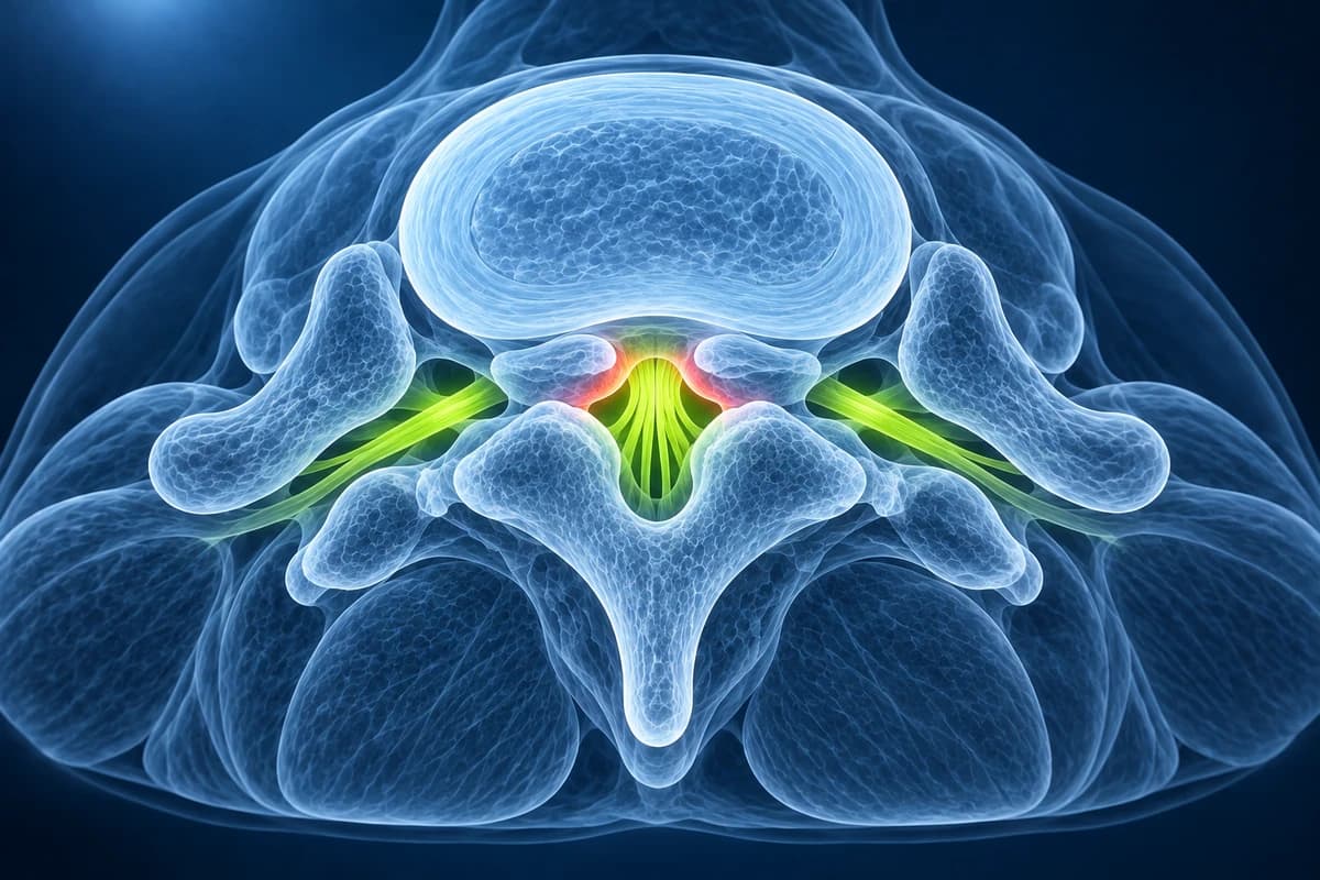
What Is Spinal Stenosis?
Foraminal Narrowing on Your MRI Report: Mild, Moderate or Severe
Foraminal narrowing means the side opening where a nerve root leaves the spine, the neural foramen, is tighter than usual. Reports grade it mild, moderate or severe by how much of the fat that cushions the nerve has disappeared on side-view images. It becomes common with age and matters most when it matches your symptoms.
Phrases you may find in the report
- “Mild left neural foraminal narrowing at L4–L5”
- “Moderate bilateral foraminal stenosis”
- “Severe right foraminal stenosis with compression of the exiting nerve root”
- “Foraminal narrowing from disc bulge and facet hypertrophy”
- “Neural foramina patent”, meaning no narrowing
What mild, moderate and severe mean
A widely used MRI grading looks at the foramen on sagittal images. Grade 0 is normal. Grade 1, mild, means the fat around the nerve is lost on two opposite sides, top and bottom or front and back. Grade 2, moderate, means fat is lost on all four sides but the nerve keeps its shape. Grade 3, severe, means the nerve root looks flattened or collapsed. Two radiologists using this scale agreed almost perfectly in the original study.
The report usually names the cause too: a bulging disc, bony spurs, a thickened ligament or enlarged facet joints, described on the facet joint syndrome page. Degenerative changes like these are frequent in people who have no back pain at all.
When foraminal narrowing changes the plan
Stenosis is diagnosed from the story of the symptoms, typically leg pain that comes on when standing or walking and eases when bending forward, and then confirmed on MRI or CT. First-line care is activity changes, pain relief and physical therapy. Surgery to widen the space is an option for selected people whose pain and limits continue despite that care, and the level that matches the symptoms guides it.
Questions about foraminal narrowing for your doctor
- Which level and side are narrowed, and which nerve root passes through there?
- Does that nerve explain where I feel pain, numbness or weakness?
- Is the narrowing caused by the disc, the facet joint or a thickened ligament?
- What would make surgery worth discussing in my case?
What Spinal Stenosis Looks Like on MRI
- Central canal narrowing with AP diameter measurement
- Lateral recess stenosis with nerve root compression
- Foraminal stenosis with nerve root impingement
- Ligamentum flavum hypertrophy contributing to stenosis
- Facet joint hypertrophy and synovial cysts
- Disc bulging as a component of stenosis
- Spinal cord compression with or without T2 signal changes (myelopathy)
Frequently Asked Questions
How is the severity of spinal stenosis measured on MRI?
The Schizas system grades central canal stenosis using dural sac morphology on MRI. Foraminal stenosis has a separate Lee grading system. Imaging severity must be interpreted alongside symptoms.
What MRI signal changes indicate active neurological compromise in stenosis?
Modic type I changes in adjacent endplates (low T1, high T2 signal indicating bone marrow edema and active inflammation) correlate with symptomatic axial pain superimposed on stenotic symptoms. Pfirrmann grade IV–V disc degeneration at the stenotic level confirms chronic degenerative etiology. High-intensity zones (HIZ) in the annulus represent annular fissuring. On axial T2 images, loss of the bright CSF rim around the dural sac and nerve root clumping indicate functionally significant compression. Myelopathy in cervical stenosis is suggested by T2 hyperintensity within the spinal cord (myelomalacia), a poor prognostic sign.
When is surgery indicated for spinal stenosis?
In the SPORT study, surgically treated patients with symptomatic spinal stenosis maintained greater improvements in pain and function than nonoperatively treated patients through four years.
Sources
- Schizas C, Theumann N, Burn A, et al. Qualitative grading of severity of lumbar spinal stenosis based on the morphology of the dural sac on magnetic resonance images. Spine (Phila Pa 1976). 2010;35(21):1919-1924.
- Lee S, Lee JW, Yeom JS, et al. A practical MRI grading system for lumbar foraminal stenosis. AJR Am J Roentgenol. 2010;194(4):1095-1098.
- Katz JN, Zimmerman ZE, Mass H, Makhni MC. Diagnosis and management of lumbar spinal stenosis: a review. JAMA. 2022;327(17):1688-1699.
- Brinjikji W, Luetmer PH, Comstock B, et al. Systematic literature review of imaging features of spinal degeneration in asymptomatic populations. AJNR Am J Neuroradiol. 2015;36(4):811-816.
- Weinstein JN, Tosteson TD, Lurie JD, et al. Surgical versus nonoperative treatment for lumbar spinal stenosis four-year results of the Spine Patient Outcomes Research Trial. Spine (Phila Pa 1976). 2010;35(14):1329-1338.
- Kreiner DS, Shaffer WO, Baisden JL, et al. An evidence-based clinical guideline for the diagnosis and treatment of degenerative lumbar spinal stenosis (update). Spine J. 2013;13(7):734-743.
Related Articles
Compare CT and MRI scans, their strengths for different conditions, radiation exposure, cost, and when each is the better choice.
Disc bulge vs protrusion vs extrusion vs sequestration, facet syndrome, vertebral fractures and stenosis — anatomy, MRI findings, and treatment options.
Pfirrmann grading of lumbar disc degeneration on MRI — Grade I (healthy) through V (severe collapse), what each grade means for symptoms and treatment.
Understand your imaging before your next appointment
MRI, X-ray or CT from a CD, folder or ZIP. A PDF report or photos of the films work too.
Free to open and preview, no account needed. An optional AI report from $10 explains your images in plain language, with questions for your doctor.
Your raw files stay on this device. Only rendered images and minimal context are sent when you request an AI report.
Medical Disclaimer: This page is for informational and educational purposes only. It does not constitute medical advice, diagnosis, or treatment. AI-generated analysis may contain errors. Always consult a qualified healthcare professional for medical decisions. Full Disclaimer