
Facet Joint Syndrome Imaging Signs: Facet Arthropathy, Effusion, and Foraminal Narrowing
Facet joint syndrome is a common cause of axial back and neck pain resulting from degenerative changes in the zygapophyseal (facet) joints. These synovial joints bear approximately 20% of the spinal axial load and are susceptible to osteoarthritis, particularly at the L4-L5 and L5-S1 levels. Facet arthropathy can produce localized pain, referred pain patterns, and secondary foraminal stenosis from hypertrophic changes. MRI and CT provide complementary information about facet degeneration.
Common Symptoms
- Axial back or neck pain that is typically unilateral and worsens with spinal extension and rotation toward the affected side
- Pain referred in a non-dermatomal pattern to the buttock and posterior thigh (lumbar) or shoulder and periscapular region (cervical) without radiculopathy
- Morning stiffness that improves with gentle movement and worsens after prolonged static postures
- Tenderness on deep palpation over the facet joints one to two finger-breadths lateral to the midline
- Pain relief in a flexed, forward-leaning posture such as pushing a shopping cart (pseudoclaudication variant in associated stenosis)
- Absence of neurological deficits on examination unless coexisting foraminal stenosis is present
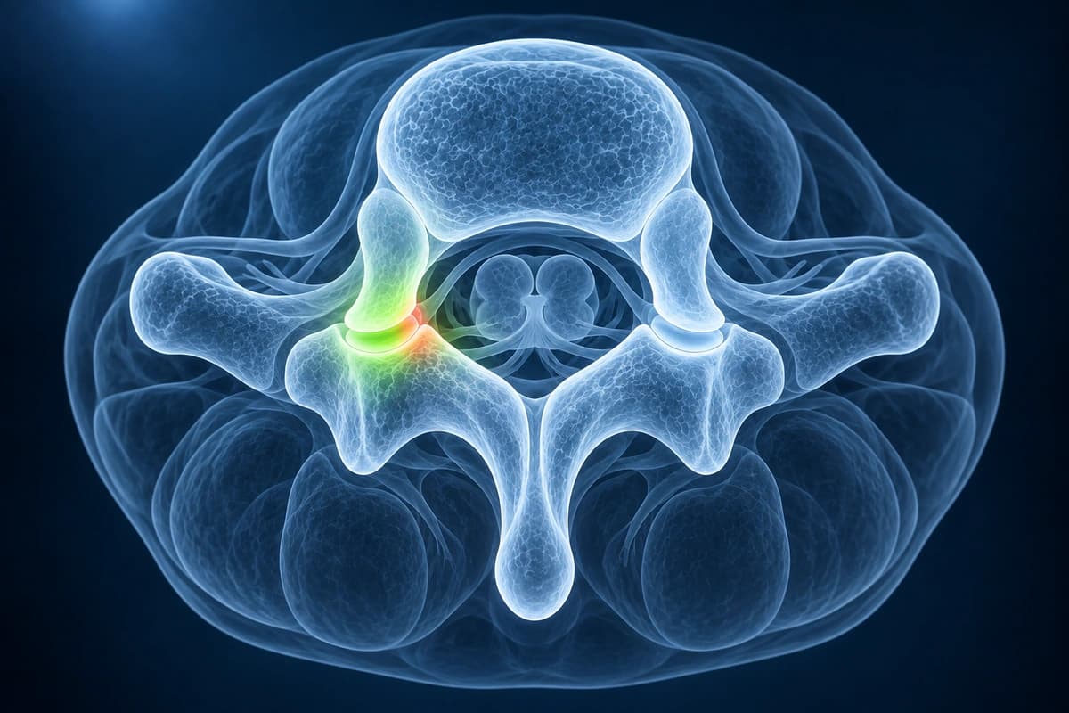
MRI is useful for facet effusion, marrow edema, synovial cysts, and adjacent nerve compromise, while CT is stronger for osteophytes, sclerosis, and pars or posterior element bone detail. If pain travels in a dermatomal pattern, compare the images with herniated disc; if sacroiliac edema or inflammatory back pain is present, compare with ankylosing spondylitis instead. A clinician should review whether imaging findings match the pain pattern before any injection or ablation plan.
Facet Hypertrophy on an MRI Report: What It Means
Facet hypertrophy means the paired joints at the back of the spine, the facet joints, have enlarged with wear. It is a sign of facet joint osteoarthritis and usually comes with joint-space narrowing and bony spurs. It can crowd the space for nerves, but on its own it does not prove that these joints are the source of your pain.
Typical facet wording in spine reports
- “Mild bilateral facet hypertrophy at L4–L5”
- “Facet arthropathy” or “degenerative facet changes”
- “Hypertrophic facet arthropathy with a small joint effusion”
- “Facet and ligamentum flavum hypertrophy narrowing the lateral recess”
How mild, moderate and severe facet hypertrophy are graded
A common four-step scale runs from grade 0, a normal joint, to grade 3. Grade 1 adds narrowing, small spurs or mild enlargement of the joint processes; grade 2 moderate spurs or enlargement with mild bone erosions; grade 3 large spurs, severe enlargement, erosions or small cysts under the joint surface. CT shows the bony change best, but in the study that compared them MRI and CT agreed within one grade in 95 to 97% of joints, so an extra CT is not needed just to grade the facets.
When facet hypertrophy matters
Facet wear becomes more common with age and with degeneration of the neighbouring disc. It matters most when the enlarged joint narrows the space for a nerve root, described on the spinal stenosis page. Whether the facets cause the pain is a clinical question: NICE advises that imaging is not needed before radiofrequency denervation, which is offered only after a positive diagnostic medial branch block.
What to ask about facet hypertrophy
- Is it mild, moderate or severe, and on which side?
- Does it narrow the space for a nerve root?
- Is there also a joint effusion or a synovial cyst?
- Would a diagnostic block help show whether these joints cause my pain?
What We Look For on Imaging
- Facet joint hypertrophy with osteophyte formation and joint space narrowing
- Facet joint effusion indicating active inflammation or instability
- Synovial cyst arising from the facet joint capsule compressing the thecal sac
- Subchondral sclerosis and erosive changes within the articular surfaces
- CT-demonstrated osteophytes, sclerosis, or vacuum phenomenon when bony detail clarifies severity
- Foraminal stenosis resulting from hypertrophic facet encroachment
- Ligamentum flavum thickening associated with posterior element degeneration
Frequently Asked Questions
How is facet joint syndrome diagnosed on imaging?
Imaging can show facet degeneration, but cannot by itself establish that a facet joint is painful. Clinical assessment and medial branch blocks inform evaluation. The cited 2020 lumbar consensus recommends a single prognostic block with more than 50% pain relief in clinical practice; other guidelines and payer policies may require higher thresholds or two blocks.
What is medial branch radiofrequency ablation and how effective is it?
Radiofrequency ablation uses heat to interrupt pain transmission through medial branch nerves. The cited 2020 lumbar consensus concludes that it may benefit well-selected patients, with medial branch blocks helping predict response. Relief is not guaranteed or permanent. Selection thresholds and the number of blocks differ across guidelines and payer policies; suitability requires clinical assessment.
How does facet joint syndrome differ from sacroiliac joint dysfunction?
In one study, three or more positive tests from a six-test sacroiliac provocation cluster had 94% sensitivity and 78% specificity compared with diagnostic injection.
Can I use a spine MRI viewer to check facet arthropathy?
Yes. A spine MRI viewer can help you inspect axial and sagittal sequences for facet joint fluid, cysts, stenosis, and asymmetric degeneration before a clinician visit. MRI viewer findings should be treated as orientation only: facet pain is confirmed by clinical examination and diagnostic medial branch blocks, not by imaging appearance alone.
Sources
- Weishaupt D, Zanetti M, Boos N, Hodler J. MR imaging and CT in osteoarthritis of the lumbar facet joints. Skeletal Radiol. 1999;28(4):215-219.
- Kalichman L, Hunter DJ. Lumbar facet joint osteoarthritis: a review. Semin Arthritis Rheum. 2007;37(2):69-80.
- Cohen SP, Raja SN. Pathogenesis, diagnosis, and treatment of lumbar zygapophysial (facet) joint pain. Anesthesiology. 2007;106(3):591-614.
- Laslett M, Aprill CN, McDonald B, Young SB. Diagnosis of sacroiliac joint pain: validity of individual provocation tests and composites of tests. Man Ther. 2005;10(3):207-218.
- Cohen SP, Bhaskar A, Bhatia A, et al. Consensus practice guidelines on interventions for lumbar facet joint pain from a multispecialty, international working group. Reg Anesth Pain Med. 2020;45(6):424-467.
- National Institute for Health and Care Excellence. Low back pain and sciatica in over 16s: assessment and management (NG59).
Understand your imaging before your next appointment
MRI, X-ray or CT from a CD, folder or ZIP. A PDF report or photos of the films work too.
Free to open and preview, no account needed. An optional AI report from $10 explains your images in plain language, with questions for your doctor.
Your raw files stay on this device. Only rendered images and minimal context are sent when you request an AI report.
Medical Disclaimer: This page is for informational and educational purposes only. It does not constitute medical advice, diagnosis, or treatment. AI-generated analysis may contain errors. Always consult a qualified healthcare professional for medical decisions. Full Disclaimer