
Sciatica on MRI & X-ray: Imaging Findings
Sciatica refers to pain radiating along the sciatic nerve, typically caused by compression or irritation of the L4, L5, or S1 nerve roots. The most common cause is a lumbar disc herniation, though foraminal stenosis, piriformis syndrome, and spondylolisthesis can also produce sciatic symptoms. MRI is the primary imaging modality for identifying the site and cause of nerve root compression. AI can help explain report terms about discs and nerves, but it cannot pinpoint the cause of your symptoms or assess their severity. A clinician must compare the scan with your symptoms and examination.
Common Symptoms
- Shooting or burning pain radiating from the low back through the buttock and down the posterior or lateral leg, following a dermatomal pattern
- Numbness and tingling in the foot or toes corresponding to the affected nerve root — lateral foot for S1, dorsum for L5, medial shin for L4
- Muscle weakness in the leg such as difficulty dorsiflexing the foot (L4–L5) or plantar flexing (S1)
- Positive straight-leg raise test reproducing radicular pain below the knee at less than 60° of hip flexion
- Pain worsened by prolonged sitting, driving, and Valsalva maneuvers
- Diminished or absent deep tendon reflex at the knee (L4) or ankle (S1) on the affected side
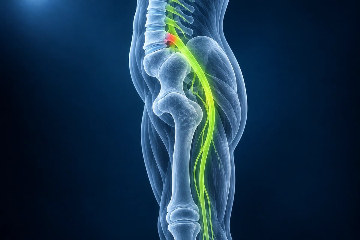
What Is Sciatica?
Do You Need an MRI for Sciatica?
Usually not at first. Guidelines advise against routine imaging for new low back pain with or without sciatica, because uncomplicated cases tend to settle on their own. MRI is considered when symptoms have not improved after about six weeks of treatment, when red flags such as bladder changes or worsening weakness appear, or when the result would change treatment.
When imaging is needed, MRI is the usual test because it shows the disc, the nerve roots and any narrowing around them. Disc bulges and protrusions are also common in people without pain, so the scan is compared with where your leg hurts and what the examination shows. For the most common causes, see herniated disc and spinal stenosis.
What Can Cause Sciatica on an MRI?
- Disc herniation contacting, displacing, or compressing the traversing or exiting nerve root
- Foraminal stenosis narrowing the neural foramen and encroaching on the exiting nerve
- Lateral recess stenosis from facet hypertrophy or ligamentum flavum thickening
- Nerve root enhancement on post-contrast sequences indicating inflammation
- Perineural cyst or mass along the sciatic nerve pathway
- Degree of thecal sac effacement and central canal narrowing at the affected level
Frequently Asked Questions
Which dermatomes are most clinically relevant in sciatica?
The sciatic nerve is formed by the L4–S3 nerve roots. Clinically, the most commonly compressed roots are L4 (medial lower leg, knee extension weakness, diminished patellar reflex), L5 (dorsal foot and great toe, extensor hallucis longus weakness, no reliable reflex loss), and S1 (lateral foot and small toe, plantar flexion weakness, diminished Achilles reflex). Dermatomal mapping guides which level on MRI to prioritize. The femoral nerve (L2–L4) is not part of the sciatic nerve but can cause similar anterior thigh pain — a positive femoral stretch test (pain with hip extension while prone) implicates L2–L4 root compression, distinguishing it from true sciatica.
Is sciatica a diagnosis or a symptom?
Sciatica is a symptom complex — radicular pain in a sciatic nerve distribution — rather than a standalone diagnosis. The most common underlying cause is a posterolateral lumbar disc herniation (contributing to 85–90% of cases), where the nucleus pulposus mechanically compresses and chemically irritates the nerve root. Other causes include foraminal stenosis from osteophytes, piriformis syndrome (extraspinal), sacroiliac joint pathology, lumbar facet synovial cysts, epidural hematoma, or neoplasm. MRI of the lumbar spine is the imaging standard to identify the structural cause and confirm the level. When MRI shows no compressive lesion, electromyography (EMG) and nerve conduction studies help localize pathology and exclude peripheral neuropathy or plexopathy.
What are the cauda equina red flags that require emergency evaluation?
Cauda equina syndrome is a surgical emergency arising from compression of the sacral nerve roots (S2–S5) below the L1 conus medullaris. Red flags include new bilateral leg weakness or sciatica, saddle anaesthesia (numbness of the perineum, inner thighs, and genitalia in an S3–S5 distribution), and dysfunction of the bladder (urinary retention with overflow incontinence being most sensitive), bowel (faecal incontinence), or sexual organs. Any patient presenting with sciatica plus bladder or bowel symptoms requires same-day emergency MRI and urgent surgical decompression — typically within 24–48 hours of symptom onset — to maximize neurological recovery.
Sources
- Jensen RK, Kongsted A, Kjaer P, Koes B. Diagnosis and treatment of sciatica. BMJ. 2019;367:l6273.
- Valat JP, Genevay S, Marty M, et al. Sciatica. Best Pract Res Clin Rheumatol. 2010;24(2):241-252.
- Brinjikji W, Luetmer PH, Comstock B, et al. Systematic literature review of imaging features of spinal degeneration in asymptomatic populations. AJNR Am J Neuroradiol. 2015;36(4):811-816.
- Gardner A, Gardner E, Morley T. Cauda equina syndrome: a review of the current clinical and medico-legal position. Eur Spine J. 2011;20(5):690-697.
- Expert Panel on Neurological Imaging; Hutchins TA, Peckham M, Shah LM, et al. ACR Appropriateness Criteria Low Back Pain: 2021 Update. J Am Coll Radiol. 2021;18(11S):S361-S379.
- Chou R, Qaseem A, Owens DK, Shekelle P. Diagnostic imaging for low back pain: advice for high-value health care from the American College of Physicians. Ann Intern Med. 2011;154(3):181-189.
- National Institute for Health and Care Excellence. Low back pain and sciatica in over 16s: assessment and management (NG59).
- National Health Service. Sciatica. NHS.
Related Articles
Discover the natural healing process of herniated discs, resorption rates, and when intervention is needed.
Safe and effective exercises for herniated disc recovery, including McKenzie method, core stabilization, and movements to avoid.
Learn when 3T spine MRI can help for subtle nerve, cord, inflammatory, or post-surgical questions, and when routine 1.5T spine MRI is usually enough.
Understand your imaging before your next appointment
MRI, X-ray or CT from a CD, folder or ZIP. A PDF report or photos of the films work too.
Free to open and preview, no account needed. An optional AI report from $10 explains your images in plain language, with questions for your doctor.
Your raw files stay on this device. Only rendered images and minimal context are sent when you request an AI report.
Medical Disclaimer: This page is for informational and educational purposes only. It does not constitute medical advice, diagnosis, or treatment. AI-generated analysis may contain errors. Always consult a qualified healthcare professional for medical decisions. Full Disclaimer